

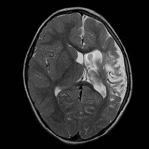
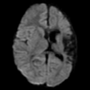

There is presence of hemi-cerebral volume loss on left side. It is involving predominantly frontal, parietal and temporal lobes. Left lateral ventricle is dilated with shift of falx to left.
There is no other focal area of abnormal signal intensity in the cerebral or cerebellar hemispheres. The grey-white matter differentiation is well maintained.
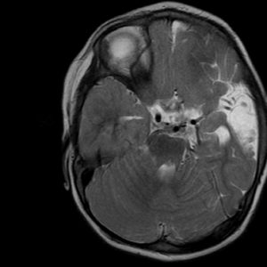
The basal ganglia, thalami, brainstem and cerebellum appear normal.
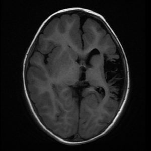
No evidence of any intracranial space occupying lesion.
Major intracranial arteries reveal normal flow void.
Paranasal sinuses appear normal.
Findings are s/o left hemi-cerebral atrophy – p/o sequel of perinatal asphyxic insult.